
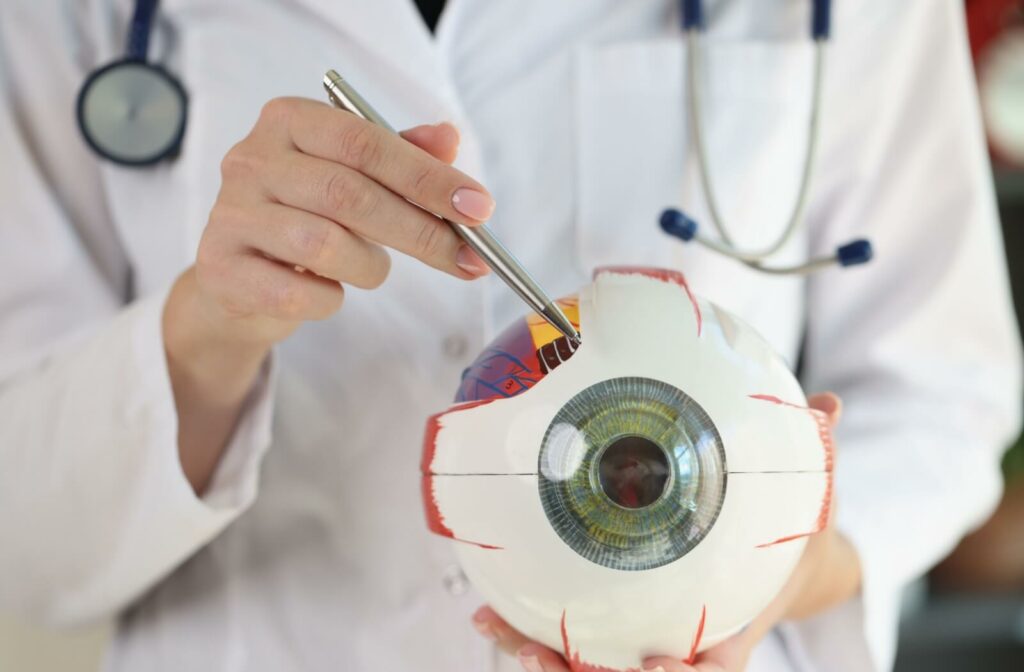
Not only are our eyes a window to our soul, but they also provide a glimpse of our overall health status. Therefore, routine eye exams are key for maintaining good ocular health and vision.
While many see eye exams merely as a way to update their vision prescription, these check-ups are necessary for detecting any abnormal shifts in the health of your eyes. Surprisingly, many eye conditions can develop silently, manifesting changes in the eye without presenting notable symptoms.
Eye diseases that can be detected during a routine eye exam include, but aren’t limited to:
- Cataracts
- Diabetic Retinopathy
- Glaucoma
- Macular Degeneration
- Retinal Detachments & Tears
Eye Diseases that can be Detected During a Routine Exam
Understandably, the onset of ocular conditions and diseases can be detected during routine eye exams. However, several general health conditions – that remain unmanaged or undetected– can also manifest signs in our eyes.
Unfortunately, not all ocular diseases can be fully cured, but many can be effectively managed when detected early. Visiting your optometrist for routine exams helps monitor the status of your ocular health and identify any possible abnormalities for timely management.
Cataracts
Cataracts develop when the eye’s natural lens becomes cloudy, leading to a progressive decline in vision. As cataracts progress, they scatter light as it enters the eye, resulting in blurred or hazy vision.
The good news is that cataracts can be effectively treated. When cataracts interfere with one’s quality of life, cataract surgery becomes a viable option to restore vision.
Cataracts are detected during a routine eye exam through several tests:
- A visual acuity test measures how well one can see at different distances, when cataracts advance, vision will become blurry and hazy.
- A slit-lamp examination allows your optometrist to examine the eye’s structure in detail, identifying any cloudiness in the lens.
- A dilated eye exam evaluates the lens and other parts of the eye for signs of cataract formation.
Diabetic Retinopathy
Diabetic retinopathy is a common complication of diabetes that specifically affects the blood vessels of the retina, responsible for capturing visual images and sending them to the brain.
This develops when high blood sugar levels damage these retinal blood vessels, causing them to leak, swell, or even close off, potentially leading to partial or total vision loss if left unmanaged. This can also put individuals at risk for retinal detachments or bleeding.
Unfortunately, there is no cure for diabetic retinopathy. However, early detection allows for better management, helping to control its progression and minimize vision damage.
Detecting diabetic retinopathy typically includes a dilated eye exam, allowing for a closer retinal examination.
Once dilated, a fluorescein angiography may be performed, where a dye is injected into the bloodstream to highlight retinal blood vessels and identify any abnormalities.

Glaucoma
Glaucoma is a group of eye diseases characterized by damage to the optic nerve, often due to increased eye pressure.
It’s considered a serious eye condition because it progresses without noticeable symptoms until the disease advances and some vision loss has occurred. Vision loss typically starts with peripheral vision and advances to central vision, severely affecting day-to-day functioning.
Glaucoma cannot be cured, However, its progression can be managed to prevent further damage, highlighting the importance of early detection.
Several tests help detect the onset of glaucoma during a routine exam:
- Tonometry helps measure eye pressure.
- An ophthalmoscope is used to examine the optic nerve for any signs of damage.
- A gonioscopy examines the eye’s drainage angle and helps determine which type of glaucoma is present.
- A visual field test assesses peripheral vision to detect any blind spots or areas of vision loss,
- OCT scans take cross-section pictures of the retina, to look for any changes indicative of glaucoma.
Macular Degeneration
Macular degeneration primarily affects the macula, the central part of the retina responsible for detailed vision such as reading and recognizing faces.
As this condition progresses, it drastically impacts one’s central visual field, causing blurriness or gaps in vision. This central vision loss makes it challenging to perform daily activities that require sharp vision, like driving or watching TV, but, peripheral vision typically remains unaffected.
Currently, there is no cure for macular degeneration, but it can be managed to slow its progression.
During the eye exam, the Amsler Grid Test is used to detect this condition. Patients look at a grid of horizontal and vertical lines, and if the lines appear wavy or have missing sections, it could indicate macular issues.
Additionally, a comprehensive dilation exam allows your optometrist to observe any signs of macular degeneration and differentiate between its dry and wet forms.
Retinal Detachments & Tears
Retinal detachments and retinal tears are serious eye conditions that require immediate medical attention.
A retinal detachment occurs when the retina, separates from its underlying support tissue. This prevents the retina from functioning properly, potentially leading to permanent vision loss if not promptly treated. A retinal tear, on the other hand, is a rip or hole in the retina that can lead to detachment.
Symptoms that could indicate retinal complications include the sudden onset of floaters, flashes of light, or a sudden shadow effect over part of the visual field.
The good news is that treatment for retinal detachments and tears is often highly effective if undertaken quickly. Laser therapies and if necessary, surgical interventions help to reattach the retina and restore vision.
Instruments including a slit lamp and ophthalmoscope allow optometrists to thoroughly examine the retina for any abnormalities while diagnostic imaging like OCT scans and retinal photos provide a detailed look at the structure of the retina.
Schedule a Visit
Similar to visiting your family doctor for an annual physical exam, these routine visits with your optometrist evaluate your overall health and identify potential issues before they escalate.
Connect with our team at Total Focus Optometry to schedule an appointment for your routine eye exam.